
When teeth don't fit together correctly, the problem is not always in the teeth.
In certain patients there is a discrepancy between the size, shape or position of the upper jaw and the lower jaw. In these cases, moving only the teeth through orthodontics can partially improve the bite, but cannot correct the skeletal origin of the alteration.
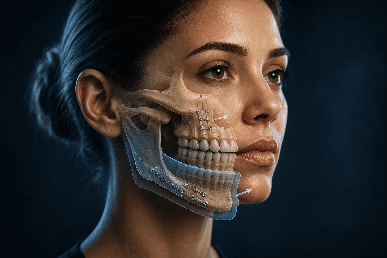
Orthognathic surgery is the set of surgical procedures aimed at repositioning the jaw bones to restore an appropriate relationship between the upper jaw, the lower jaw, the teeth and the rest of the facial structures.
Its objective is not simply to achieve a particular facial appearance. The treatment seeks to achieve a balance between occlusion, function, stability and facial harmony.
The relevant question, therefore, is not only whether a patient has "a protruding jaw" or "a receded chin", but rather which component of their craniofacial structure is altered, in what dimension, and what consequences that alteration produces.
What exactly is orthognathic surgery?
The term orthognathic comes from the Greek words orthos, correct, and gnathos, jaw.
In clinical practice it is used to describe interventions in which the upper jaw, the lower jaw, or both are surgically mobilized to modify their three-dimensional position.
Depending on the existing deformity, it may be necessary to:
- advance, impact, lower or reposition the upper jaw;
- advance or set back the lower jaw;
- correct lateral deviations;
- modify certain vertical or transverse discrepancies;
- operate on both jaws in a bimaxillary surgery;
- combine, when indicated, complementary procedures such as a genioplasty.
Modern orthognathic surgery is usually performed through intraoral approaches. Once the planned osteotomies have been carried out, the bone segments are moved to the previously determined position and stabilized using internal fixation systems, generally titanium plates and screws.
But the operation is only part of the treatment. In most patients there is a coordinated process between oral and maxillofacial surgery and orthodontics, because the position of the teeth must be compatible with the new position of the jaw bones.
The fundamental concept: a malocclusion can be dental or skeletal
This difference is probably one of the keys to understanding when orthognathic surgery may be necessary.
Some bite alterations are fundamentally due to the position of the teeth. In these patients, orthodontics can correct the problem by moving the teeth within their bone bases.
In other cases, however, there is a discrepancy between the skeletal bases themselves.
For example, a patient may present an anterior crossbite because:
- the incisors are poorly positioned;
- the upper jaw is insufficiently developed;
- the lower jaw has excessive projection;
- there is simultaneously maxillary retrusion and mandibular prominence.
Although these situations may clinically appear similar, they do not represent the same diagnosis and should not be treated in the same way.
Orthognathic surgery is especially indicated when the magnitude or nature of the skeletal discrepancy exceeds what can reasonably and stably be corrected through dental movements alone.
When is orthognathic surgery indicated?
There is no single measurement that on its own determines the need for surgery.
The indication is established after integrating the facial examination, the relationship between the jaws, the occlusion, the position of the teeth, growth, function, radiological images and the patient's expectations.
Among the situations that most frequently may justify an orthognathic assessment are the following.
Mandibular prognathism
Mandibular prognathism describes a situation in which the lower jaw is excessively advanced in relation to the upper jaw and the rest of the face.
It can produce a skeletal class III relationship, an anterior crossbite and a characteristic facial profile.
However, a class III does not necessarily mean that the jaw is excessively large. In some patients the main problem is a deficiency in growth of the upper jaw.
Distinguishing between the two situations is essential because the surgical treatment can be completely different.
Retrognathia or mandibular deficiency
In other patients the lower jaw is positioned excessively behind the upper jaw.
This can generate a significant anteroposterior discrepancy, increased dental overjet, difficulty achieving passive lip closure, and alterations in the balance of the lower third of the face.
When the discrepancy is predominantly skeletal, mandibular advancement may be part of the treatment.
In selected patients, mandibular and maxillomandibular position may also have implications for airway dimensions, an issue that must be analyzed individually and within the full clinical context.
Maxillary hypoplasia or retrusion
An insufficiently projected upper jaw can alter the dental relationship, upper lip support and the architecture of the midfacial third.
In certain class III deformities, advancing the upper jaw allows simultaneous correction of the skeletal relationship and the occlusion.
The diagnosis must determine how much is attributable to the maxilla, how much to the mandible, and how much to the compensatory position of the teeth.
Skeletal open bite
In an open bite, certain upper and lower teeth do not make contact when the patient closes the mouth.
Open bites of predominantly dentoalveolar origin may respond to orthodontic treatment.
Severe skeletal forms are different. They may be associated with a vertical growth pattern, alterations in maxillary and mandibular position, increased lower facial height and certain dental compensations.
When the skeletal component is significant, modifying only the inclination of the teeth may not adequately resolve the structural problem.
Facial asymmetry and laterognathism
A human face is never perfectly symmetrical, and small differences between the two sides are normal.
The situation changes when there is a clinically significant deviation of the maxilla, the mandible, the chin or the dental midlines.
Asymmetries can have multiple origins: growth alterations, condylar hyperplasia, traumatic sequelae, congenital anomalies or developmental differences between both sides of the jaw.
In these cases it is especially important to determine whether the deformity is stable or whether an active growth process persists before planning its definitive correction.
Vertical maxillary excess
Some patients present excessive vertical development of the upper jaw.
It can manifest as significant gingival exposure when smiling, lip incompetence, increased lower facial height, or certain open bites.
When the cause is fundamentally skeletal, maxillary impaction through osteotomy can modify both the dental position and the facial proportions.
Transverse deficiencies and crossbites
The upper jaw can also be narrow in relation to the lower jaw.
Depending on the patient's age, the magnitude of the discrepancy and its anatomical characteristics, there are different strategies for orthodontic or surgically assisted expansion.
Not all crossbites require conventional orthognathic surgery, but a significant transverse discrepancy must form part of the three-dimensional analysis of the case.
What problems can orthognathic surgery correct?
The most evident consequence of repositioning the jaws is a modification of the relationship between the teeth, but limiting the treatment to "correcting the bite" is incomplete.
A dentofacial deformity can simultaneously affect different levels.
Occlusion
The goal is to achieve a functional and stable relationship between the dental arches.
This can improve problems such as:
- crossbite;
- open bite;
- large anteroposterior discrepancies;
- certain crossbites;
- significant deviations of the midlines.
Chewing
A poor relationship between the jaws can reduce the efficiency with which the teeth contact and process food.
The available evidence shows long-term improvements in different dimensions of oral function and quality of life after orthognathic treatment, although a temporary decrease in function is expected during the early postoperative stages.
Lip competence and soft-tissue balance
The position of the bones largely determines the support of the lips, cheeks and chin.
For this reason, any skeletal movement also produces a response from the soft tissues.
Planning must anticipate these changes. It is not just a bone segment that is moved: a three-dimensional facial architecture is modified.
Facial symmetry
When there is a significant skeletal deviation, surgery can reposition the jaws relative to the facial midline and improve the balance between the two sides of the face.
In complex cases, asymmetries require especially rigorous three-dimensional planning.
Airway in selected patients
The relationship between craniofacial structure and the airway has gained particular relevance in recent decades.
In certain patients with obstructive sleep apnea syndrome, maxillomandibular advancement constitutes a specific surgical strategy. However, this does not mean that any orthognathic surgery automatically improves breathing, nor that it should be indicated for that purpose without an adequate study.
Airway evaluation, when clinically justified, must be integrated with the facial, occlusal and functional diagnosis.
Orthognathic surgery and facial aesthetics: an inseparable but not identical relationship
There is some confusion in presenting orthognathic surgery as an exclusively functional intervention or, at the opposite extreme, as cosmetic surgery.
Neither description is sufficiently accurate.
Changing the position of the upper or lower jaw inevitably modifies the face.
A maxillary advancement can change the projection of the midface and lip support. A maxillary impaction can alter dental and gingival exposure. A mandibular movement modifies the contour of the lower third, and correcting an asymmetry changes the relationships between the different facial lines.
Therefore, function and facial morphology must be planned together.
The goal is not to pursue universal mathematical proportions or a particular facial model, but to find an anatomically coherent solution for each patient.
How is a dentofacial deformity diagnosed?
Rigorous orthognathic planning begins long before using a virtual planning program.
It begins with diagnosis.
The evaluation must integrate several levels.
Facial examination
The analysis is performed frontally and in profile, but also in three dimensions.
Among other aspects, the following are studied:
- vertical proportions;
- symmetry;
- midlines;
- maxillary and mandibular projection;
- lip support;
- lip competence;
- dental exposure at rest and when smiling;
- chin position;
- relationships between the nose, lips and lower facial third.
Intraoral and occlusal examination
It is necessary to determine how the two arches relate to each other and which part of the alteration corresponds to the teeth and which part to the bone bases.
The so-called dentoalveolar compensations must also be identified.
When the jaws grow into discrepant positions, the teeth may spontaneously tilt in the opposite direction in an attempt to maintain some contact between them. This compensation can make the deformity appear externally smaller than it actually is.
Radiological study
Cephalometry continues to be a useful tool for analyzing craniofacial relationships.
In certain cases, especially in the presence of asymmetries or complex deformities, cone-beam computed tomography and three-dimensional reconstructions allow the anatomy to be studied with greater precision.
AO Foundation notes precisely that clinical examination, cephalometry and standardized photography are fundamental elements of the diagnosis, incorporating three-dimensional studies when the complexity of the case requires it.
Digital records and virtual planning
Digital models and virtual surgical planning allow bone movements to be simulated, anatomical interferences to be analyzed, and the plan to be transferred to the operating room using digitally designed splints or positioning systems.
Technology improves planning capability, but does not replace clinical judgment.
Software can execute a five-millimeter movement. It cannot decide on its own whether those five millimeters represent the correct movement for that patient.
Why is orthodontics usually necessary before surgery?
This is one of the questions that most surprises some patients.
Before orthognathic surgery, the teeth may be tilted in a compensatory manner due to the incorrect position of the jaws.
In order to place the bones in an appropriate relationship, those compensations must be corrected.
This process is called orthodontic decompensation.
As a result, during one phase of treatment the bite may even appear worse. This does not mean the treatment is failing. It means the teeth are being correctly positioned within each jaw so that they can later relate to each other appropriately once the skeletal movement is performed.
After surgery there is usually an additional phase of orthodontics aimed at refining the occlusion and stabilizing the result.
For this reason, orthognathic treatment should be understood as a coordinated process between specialties and not as an isolated operation.
Which surgeries are most frequently used?
The technique depends on the deformity.
Le Fort I osteotomy
This allows the upper jaw to be mobilized.
Depending on the surgical plan, its position can be advanced, impacted, lowered, rotated, or corrected transversely within certain limits.
Bilateral sagittal split osteotomy of the mandible
This is one of the most commonly used techniques for repositioning the lower jaw.
It allows advancement, setback or asymmetric corrections to be performed depending on the characteristics of the case.
Bimaxillary surgery
When the deformity simultaneously involves the maxilla and the mandible, it may be necessary to operate on both in the same procedure.
Complex three-dimensional deformities, certain asymmetries, vertical discrepancies and combinations of anteroposterior problems are situations in which bimaxillary movements frequently need to be considered.
Genioplasty
Genioplasty modifies the position or morphology of the chin.
It can be performed in isolation in certain patients or combined with orthognathic surgery when necessary to complete the balance of the lower facial third.
It is important to differentiate it from a mandibular movement: repositioning the chin and repositioning the jaw are procedures with different anatomical objectives.
Are there alternatives to orthognathic surgery?
Yes, but they depend on the diagnosis.
In mild or moderate discrepancies, compensatory orthodontic treatment may be considered under certain circumstances.
Orthodontics camouflages part of the skeletal discrepancy by modifying the position of the teeth without significantly changing the position of the bone bases.
It can be an excellent solution in the right patient.
What matters is understanding its limit: orthodontics moves teeth; orthognathic surgery modifies the relationship between the jaws.
When the deformity is significant, attempting to correct it through excessive dental movements can compromise the periodontal, occlusal, or facial result, or its stability.
The choice between orthodontic compensation and combined orthodontic-surgical treatment must be made after clearly explaining what each alternative can achieve and what aspects will remain uncorrected.
What is recovery like after orthognathic surgery?
Recovery depends on the procedures performed and the individual characteristics of each patient.
Facial swelling is expected and tends to be more intense during the first few days, decreasing progressively over the following weeks. The more subtle soft-tissue changes may continue to evolve for several months.
During the early phases, the consistency of the diet is also adapted while bone consolidation progresses.
In contemporary orthognathic surgery, the jaws are usually stabilized using internal fixation with plates and screws, and prolonged rigid intermaxillary fixation of both jaws is not the norm in many current protocols. Orthodontic elastics can indeed be used to guide the occlusion during recovery.
The return to work or school depends on the type of activity and individual progress. Rather than establishing a universal figure, it is preferable to assess each patient's recovery process.
Risks and limitations of orthognathic surgery
Every surgery carries risks, and orthognathic surgery is no exception.
The nature and probability of each complication depend on the procedure performed, the planned movement, individual anatomy and the patient's general condition.
Among the aspects that should be explained before treatment are:
- temporary or persistent alterations in sensitivity;
- infection;
- bleeding;
- problems related to fixation systems;
- occlusal alterations;
- partial relapse of the movement achieved;
- periodontal or dental problems in certain circumstances;
- exceptional need for additional procedures;
- soft-tissue aesthetic response different from expected;
- possible effects on the temporomandibular joint.
Special attention should be given to sensory alteration after certain mandibular procedures, due to the proximity of the inferior alveolar nerve.
Decreased sensitivity of the lower lip and chin is relatively common in the initial period after certain mandibular osteotomies. In many patients it improves progressively, but there is a possibility that some degree of sensory alteration may persist.
Responsible surgical indication requires discussing these risks with the same clarity used to explain the possible benefits.
Are the results stable?
The stability of orthognathic surgery depends on multiple factors:
- type of deformity;
- direction and magnitude of the movements;
- quality of the planning;
- stability of the temporomandibular joint;
- surgical technique;
- fixation;
- muscular adaptation;
- residual growth;
- orthodontic treatment;
- compliance with the retention phase.
The goal is not to have a biological anatomy remain absolutely immobile for the rest of one's life, but to obtain a clinically stable correction.
Precisely for this reason, the preoperative diagnosis must identify factors capable of compromising that stability.
Orthognathic surgery does not begin in the operating room
This may be the most important idea for understanding this treatment.
The outcome of orthognathic surgery does not depend solely on correctly performing an osteotomy.
It depends on having previously understood:
- where the deformity truly lies;
- which part is dental and which part is skeletal;
- what compensations have occurred;
- how the maxilla and mandible relate to the cranial base;
- what effect each movement will have on the occlusion;
- how the soft tissues will respond;
- whether there is an associated functional alteration;
- and what the reasonable goals are for that patient.
Surgery is the execution of a diagnosis.
And the more precise that prior interpretation is, the more coherent the treatment can be.
Frequently asked questions about orthognathic surgery
Is orthognathic surgery a cosmetic surgery?
Not exclusively. It can produce significant aesthetic changes because it changes the structure of the face, but its planning integrates skeletal, occlusal and functional aspects.
Do all patients need surgery on both jaws?
No. Some need only maxillary surgery, others mandibular surgery, and others require bimaxillary surgery. The decision depends on where the deformity is located and the three-dimensional result being pursued.
Can orthognathic surgery be done without orthodontics?
There are specific protocols in which surgery can be performed before completing orthodontic preparation, known as surgery first. These are not applicable to all patients. Case selection and coordination between the surgeon and the orthodontist are especially important.
Can surgery correct an asymmetric face?
It can correct asymmetries whose origin lies in the position or morphology of the jaws. The cause of the asymmetry must first be determined, and it must be confirmed, when relevant, that there is no active pathological growth process.
Does an incorrect bite always require surgery?
No. Many malocclusions can be treated exclusively through orthodontics. Surgery is considered when there is a skeletal component whose magnitude or characteristics make a purely dental correction insufficient.
Will my face change a lot?
It depends on the deformity and the movements required. Some treatments produce discreet modifications and others more evident changes. This is precisely why facial analysis and planning form an essential part of the process.
Does orthognathic surgery improve quality of life?
Studies overall show an improvement in various indicators of oral health-related quality of life after orthognathic treatment, especially in the medium and long term. A systematic review published in 2025 found improvements in functional aspects such as chewing and speech, although it also highlighted the need for further long-term outcome studies.
A decision that must start from the diagnosis
Two patients may present an apparently similar bite and require completely different treatments.
One may have a predominantly dental alteration. Another, a maxillary deficiency. Another, a mandibular excess. And a fourth, a combination of both problems accompanied by a vertical or transverse alteration.
For this reason, orthognathic surgery should not be indicated based on a photograph, an isolated X-ray, or a specific aesthetic concern.
It requires studying the dentofacial system as a whole.
When there is a significant skeletal deformity, correct planning makes it possible to treat not only how the teeth make contact, but also the relationship between the maxilla and mandible, function, and facial balance.
That is the true purpose of orthognathic surgery.
References and sources
- American Association of Oral and Maxillofacial Surgeons (AAOMS). Orthognathic Surgery Indications. Clinical Papers, 2025.
- AO Foundation, Surgery Reference. Planning of Orthognathic Surgery.
- Zheng L, Saddki N, Su L, Ab Rahman N. The impact of orthognathic surgery on oral function-related quality of life: a systematic review. Oral and Maxillofacial Surgery. 2025.
- Queen Victoria Hospital NHS Foundation Trust. A guide for patients considering orthodontics and jaw surgery.