
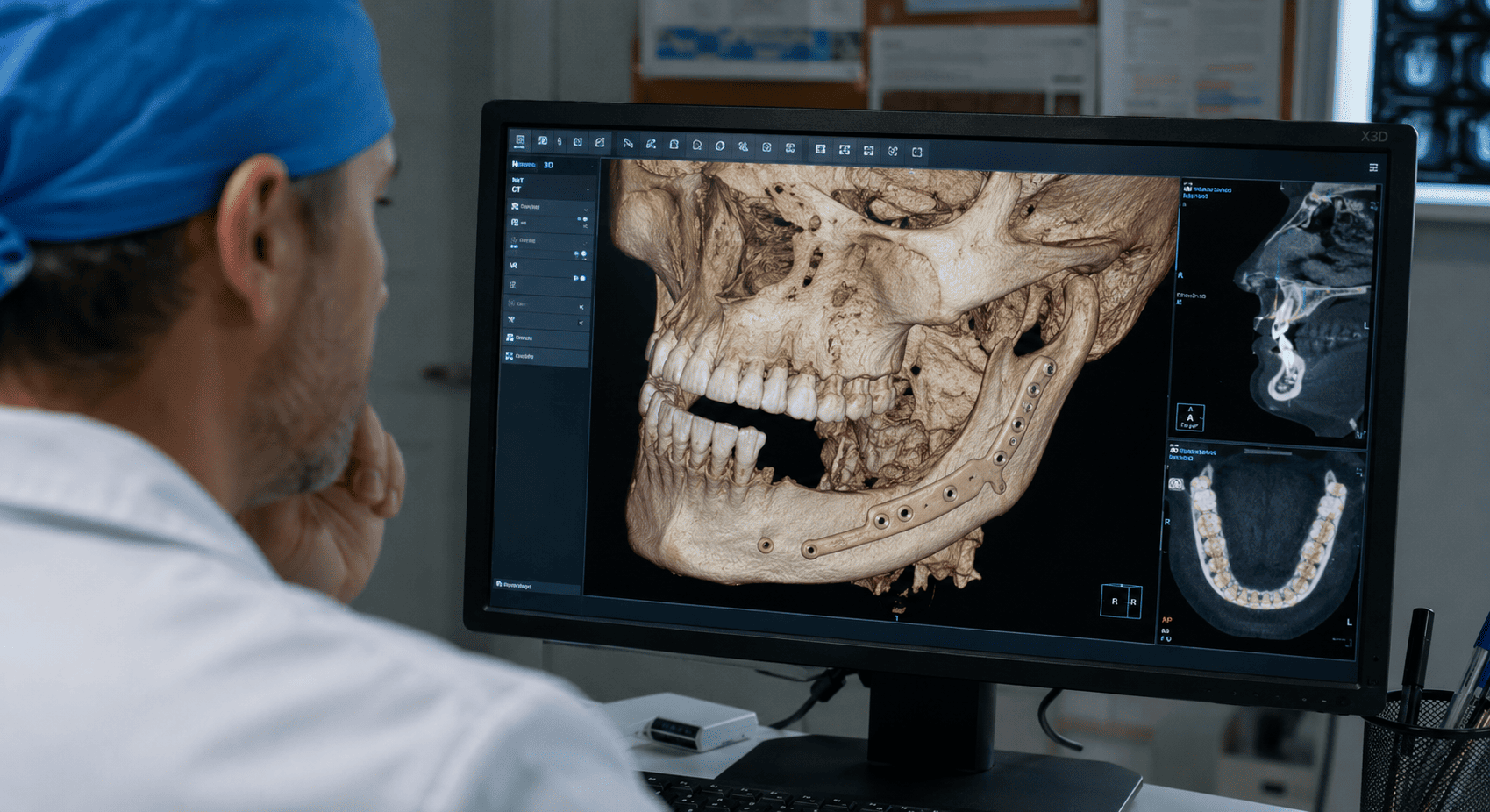
The mandible is not simply a bone that defines the lower contour of the face.
It participates in chewing, maintains the position of the lower dentition, contributes to speech and swallowing, provides attachment for numerous muscles and forms part of a functional system involving the tongue, the floor of the mouth, the facial soft tissues and the temporomandibular joints.
When a significant part of the mandible must be resected or is lost as a consequence of disease, trauma or a treatment-related complication, the reconstructive problem does not simply consist of filling a bone gap.
An attempt must be made to re-establish a structure capable of restoring continuity, form and function, providing support to the soft tissues and, when conditions allow, creating a suitable base for dental rehabilitation.
This is why mandibular reconstruction should begin before resection whenever possible.
The question is not only what to reconstruct the mandible with, but what will be lost, what must be preserved and what functions will need to be recovered.
What is mandibular reconstruction?
Mandibular reconstruction comprises different procedures aimed at repairing a defect of the mandible.
The concept includes very different situations.
A small bone defect with preserved mandibular continuity does not pose the same problem as a segmental mandibulectomy in which an entire segment of bone disappears.
Nor is reconstructing bone alone equivalent to simultaneously reconstructing the mandible, oral mucosa, floor of the mouth, tongue, skin and other soft tissues.
For this reason, before choosing a technique it is necessary to characterize the defect.
Among the fundamental questions are:
- is mandibular continuity preserved?;
- how much bone length is missing?;
- does the defect affect the anterior or lateral region?;
- does it include the angle or the mandibular ramus?;
- has the condyle been lost?;
- is there an associated mucosal or skin defect?;
- which teeth will remain?;
- has the territory received radiotherapy?;
- which recipient vessels are available?;
- is implant-supported rehabilitation planned afterward?
The answers to these questions determine much of the reconstructive strategy.
When may a mandibular reconstruction be necessary?
The causes are diverse, but there are several main scenarios.
After tumor resection
Oncologic surgery constitutes one of the most important indications.
Malignant tumors of the oral cavity, oropharynx, salivary glands or adjacent structures can infiltrate the mandible and require bone resection to achieve adequate oncologic treatment.
There are also locally aggressive benign tumors capable of producing extensive mandibular destruction and requiring segmental resections.
In these patients there is a principle that must not be reversed:
reconstruction must be adapted to the resection that is oncologically necessary; the resection must not be made insufficient in order to facilitate reconstruction.
When the resulting defect is predictable, reconstructive planning can be carried out simultaneously with planning of the resection.
Mandibular osteoradionecrosis
Radiotherapy used to treat head and neck tumors can, in certain patients, produce severe damage to the irradiated bone known as osteoradionecrosis.
Not all cases require mandibular resection.
However, when there is advanced disease with significant bone destruction, pathological fracture, fistulas, persistent infection or structural deterioration that cannot be controlled with conservative strategies, resection of the affected bone and reconstruction with vascularized tissue may be necessary.
This scenario presents specific difficulties because the recipient territory has been irradiated and tissue quality may be compromised.
Complex trauma
Common mandibular fractures can be treated by reduction and fixation without needing to reconstruct entire segments.
The problem is different with high-energy trauma involving extensive bone loss, gunshot injuries, contaminated defects or sequelae of previous trauma.
In these cases it may be necessary to reconstruct both bone continuity and the associated soft tissues.
Infections and osteonecrosis
Certain severe bone infections, refractory osteomyelitis or other necrotic processes can produce mandibular destruction that requires resection of non-viable tissue.
The reconstructive indication depends on the extent of the process, control of the infection and the patient's local and systemic condition.
Congenital defects or sequelae of previous surgeries
Some craniofacial deformities, sequelae of prior interventions or failed reconstructions may require secondary procedures.
These cases tend to be especially complex because the anatomy has been previously altered and there may be scarring, absence of suitable vessels, occlusal alterations and tissue deficits.
The defect determines the reconstruction
One of the fundamental ideas in reconstructive surgery is that not every centimeter of mandible has the same functional significance.
Losing a lateral segment is not equivalent to losing the anterior region.
The symphysis and the anterior body contribute decisively to chin projection and the mandibular arch. The lateral segments determine part of the facial contour and support the posterior dental sectors. The ramus and condyle participate in the mandible's relationship with the skull base.
Moreover, complexity increases when a defect spans several anatomical regions.
This is why classification systems exist to describe mandibular defects in a reproducible way.
The classification proposed by Brown for defects after oncologic resection uses four anatomical reference points — the two mandibular angles and the canine regions — and distinguishes lateral, hemimandibular, anterior and extensive defects, incorporating additional categories when condylar resection is present.
The importance of classification is not academic.
Correctly defining the defect allows the geometric and functional difficulty of reconstructing it to be anticipated.
Mandibular continuity: an essential distinction
From a reconstructive standpoint there is a fundamental difference between a marginal resection and a segmental one.
Marginal mandibulectomy
A portion of the mandible is removed, but sufficient bone continuity is maintained.
In certain patients a vascularized bone reconstruction is not necessary.
The strategy can focus on achieving adequate soft-tissue coverage and preserving a structurally stable mandible.
Segmental mandibulectomy
A transverse segment of the mandible is completely removed.
Continuity is lost.
This alters mandibular biomechanics and can produce displacement of the remaining segments, occlusal alterations, asymmetry, chewing difficulties and significant changes in soft-tissue support.
When the patient's general condition and oncologic status allow it, significant segmental defects usually require reconstruction capable of restoring that continuity.
What does a good mandibular reconstruction aim to recover?
Reconstructive success should not be defined solely by graft survival.
There are several objectives.
Bone continuity
Restoring a stable mandibular arch constitutes the structural basis of the reconstruction.
Facial form
The mandible contributes decisively to the width, projection and symmetry of the lower third of the face.
A geometrically deficient reconstruction can maintain bone continuity and still produce a significant facial alteration.
Occlusion
When residual dentition is present, the relationship between the reconstructed segments and the remaining teeth must be part of the planning.
A relatively small deviation in mandibular position can translate into a significant occlusal discrepancy.
Chewing
Restoring continuity does not necessarily mean restoring chewing function.
Function also depends on the dentition, the soft tissues, tongue mobility, the temporomandibular joint and the possibility of dental rehabilitation.
Speech and swallowing
In major oncologic resections, mandibular loss may be accompanied by resections of the tongue, floor of the mouth or mucosa.
In these patients, the functional outcome depends on both soft-tissue reconstruction and the bone component.
Possibility of dental rehabilitation
When it is reasonable to consider it, reconstruction should factor in from the outset where the future alveolar ridge will be located and whether the reconstructed bone will allow implant-based rehabilitation.
This point changes the way the procedure should be understood:
the optimal position of the bone for reconstructing the inferior mandibular border does not always coincide with the optimal position for reconstructing the future dentition.
Planning must attempt to resolve both needs.
Why is vascularized bone used?
In complex mandibular defects, one of the most important reconstructive tools is microvascular tissue transfer.
A vascularized free bone flap allows bone to be transferred from another region of the body together with its artery and vein.
Once placed in the mandibular defect, its vessels are connected by microsurgery to vessels in the neck.
The transferred bone thus maintains its own blood supply.
This represents a fundamental biological difference compared with a non-vascularized bone graft.
Vascularized tissues are especially important in:
- extensive segmental defects;
- the need to simultaneously reconstruct bone and soft tissues;
- previously irradiated territories;
- major oncologic resections;
- certain complex defects or secondary reconstructions.
The choice of donor tissue depends on the defect and should not be based solely on familiarity with a technique.
Free fibula flap: why it is used so frequently
The free fibula flap has become one of the main resources for mandibular reconstruction and continues to be considered the reference flap for many complex defects.
There are several reasons for this.
The fibula provides a considerable length of vascularized bone and allows multiple osteotomies to be performed to reproduce the mandibular curvature.
Its vascular pedicle is usually suitable for microsurgical reconstruction, and a skin island can be included when soft tissue needs to be reconstructed.
In addition, its bone structure allows dental implants to be considered later in selected patients.
However, describing it as the best universal solution would be incorrect.
The fibula has a specific geometry and bone height. The defect may require different characteristics, and the vascular anatomy of the lower limbs must be evaluated when relevant before using it.
Reconstruction should select the flap for the patient, not select the patient for the flap.
What other options exist?
Iliac crest flap
The iliac crest provides a significant amount of bone and a morphology that can be favorable for certain reconstructions.
Its bone height can offer advantages when future dental rehabilitation is an important goal.
It can also be combined with soft-tissue components depending on the flap design.
The choice between fibula and iliac crest cannot be reduced to an absolute superiority of one over the other. Comparative meta-analyses have not shown a significant difference in total flap loss and show different profiles of complications and reconstructive characteristics.
Scapular system
Flaps based on the subscapular system offer great versatility for combining bone components and different soft-tissue territories.
This can be especially useful when the defect is not predominantly bony and there is a significant need for three-dimensional soft-tissue reconstruction.
Its geometry, pedicle, surgical positioning and patient characteristics must be integrated into the decision.
Non-vascularized bone grafts
Non-vascularized grafts continue to have indications in certain selected defects.
Their use depends on factors such as the extent of the defect, quality of the recipient bed, absence of radiotherapy and local conditions.
They should not be considered equivalent to a vascularized flap in extensive defects or in biologically compromised territories.
Reconstruction plates
Plates can serve different functions: temporary stabilization, support for certain defects or part of specific reconstructive strategies.
But a plate is not living tissue.
In complex segmental defects, especially when radiotherapy has been given or soft-tissue coverage is deficient, a reconstruction based exclusively on fixation hardware can present problems such as exposure, infection or hardware fracture.
For this reason, reconstructing mechanical continuity and biologically reconstructing a mandible are not equivalent concepts.
Planning begins with the resection
In an oncologic reconstruction, the ablative surgeon and the reconstructive team should not work as two independent processes.
Planning begins by defining:
- which tissue must be resected;
- which structures can be safely preserved;
- what the likely geometry of the defect will be;
- which tissues will need to be replaced;
- which recipient vessels will be available;
- which reconstruction offers the best balance between morbidity and function.
In certain cases, this information can be studied through virtual planning before entering the operating room.
This makes it possible to move from an essentially reactive reconstruction — "see what defect remains and reconstruct it" — to a strategy in which an important part of the problem has already been analyzed beforehand.
3D virtual planning of mandibular reconstruction
Virtual planning has had a particularly significant impact on mandibular reconstruction.
Starting from a CT scan, the craniofacial skeleton can be reconstructed three-dimensionally.
Using that model, it is possible to simulate the planned resection and study the resulting defect.
A three-dimensional model of the donor bone, the fibula for example, can subsequently be incorporated.
The process allows planning of:
- resection margins;
- defect length;
- osteotomies of the donor bone;
- number and length of segments;
- orientation of each segment;
- restoration of the mandibular arch;
- position of the bone relative to the dentition;
- relationship with the maxilla;
- placement of fixation systems.
Cutting guides, positioning guides, printed models and, in certain cases, patient-specific plates can be derived from this planning.
Reconstructing a curve with a straight bone
This is one of the fundamental geometric problems of fibula reconstruction.
The fibula is essentially a long bone.
The mandible is a curved three-dimensional structure.
To convert one into the other, the fibula must be divided by osteotomies and its segments oriented so as to reproduce the mandibular arch.
Each osteotomy involves a decision:
- where to cut;
- how much bone to preserve;
- what angle to create;
- how to maintain the vascular pedicle;
- how to contact the segments;
- how to reproduce the mandibular shape.
A relatively straight lateral reconstruction may require a less complex geometry than an anterior reconstruction that includes the symphysis.
This is why anterior and extensive defects can pose especially significant reconstructive difficulties.
Virtual planning makes it possible to quantify these decisions before the cuts are made.
What does reconstructing with precision mean?
In reconstructive surgery, the word precision needs a definition.
It does not simply mean that the bone is positioned within a few millimeters of a digital model.
Precision must be assessed based on what matters clinically:
- continuity;
- symmetry;
- mandibular width;
- projection;
- position of the angles;
- maxillomandibular relationship;
- occlusion;
- condylar position when relevant;
- height of the reconstructed bone;
- contacts between segments;
- feasibility of future dental rehabilitation.
A model can be geometrically precise and still fail to adequately address a functional need.
Technology is useful because it allows measurement.
Reconstructive judgment determines what is worth measuring.
Occlusion cannot be a secondary consideration
When teeth remain, the reconstruction must relate to them.
If the reconstructed mandible ends up transversally displaced, rotated or with an incorrect length, the relationship with the upper maxilla can be altered.
For this reason, digital dental records and different positioning strategies can be used to preserve or recover the maxillomandibular relationship.
In certain complex reconstructions, thinking first about the external shape of the mandibular border and then trying to adapt the dentition can produce a structurally correct but prosthetically unfavorable result.
Rehabilitation begins during reconstructive planning, not after the bone has consolidated.
Reconstruction oriented toward dental rehabilitation
One of the most important conceptual changes in contemporary mandibular reconstruction consists of incorporating the future dentition into the plan.
Surviving an oncologic resection naturally remains the priority.
But when prognosis, the patient's condition and the planned treatment allow it, functional recovery should be considered from the outset.
This requires analyzing:
- bone volume and position;
- relationship with the upper arch;
- prosthetic space;
- peri-implant soft tissues;
- planned or previous radiotherapy;
- the possibility and timing of implant placement;
- design of the future prosthesis.
Implants can be placed later after consolidation or, in carefully selected patients within specific protocols, during the reconstructive procedure itself.
The decision must be individualized.
The fact that an implant can technically be placed does not mean there is always an indication to do so immediately.
Immediate or delayed reconstruction?
Whenever conditions allow, certain defects can be reconstructed in the same procedure as the resection.
This makes it possible to immediately restore mandibular continuity and avoids maintaining the anatomical consequences of a segmental defect for a prolonged period.
However, not all scenarios are the same.
Infection, general condition, extent of the trauma, uncertainty about tissue viability, previous procedures or certain oncologic circumstances may require a different strategy.
Delayed reconstruction also presents its own challenges: fibrosis, scarring, soft-tissue contracture, segment displacement and possible alterations of the recipient vessels.
For this reason, the timing of reconstruction must be part of the plan and not an isolated decision.
Soft-tissue reconstruction can be as important as bone reconstruction
A common mistake is to imagine mandibular reconstruction as an exclusively skeletal problem.
In many oncologic defects this is not true.
It may be necessary to simultaneously reconstruct:
- oral mucosa;
- floor of the mouth;
- tongue;
- skin;
- facial volume;
- coverage of the fixation hardware.
A perfectly positioned bone can fail functionally if the soft tissues are insufficient, restrictive or unable to provide adequate coverage.
For this reason, the choice of flap also depends on how much soft tissue is needed and where it must be placed.
In some patients, the need for soft tissue can influence the choice of donor site as much as the bone defect itself.
What happens with the mandibular condyle?
Defects that include the condyle add another level of complexity.
The condyle is part of the temporomandibular joint and its position influences mandibular mechanics.
When it must be resected, different reconstructive strategies exist depending on the clinical context, extent of the defect, age, etiology and available tissues.
There is no single solution applicable to all patients.
Reconstruction must consider not only continuity between the mandibular body and the skull base, but also stability, mobility, occlusion and risk of displacement.
Precisely for this reason, modern classification systems distinguish defects that include condylar loss.
The donor site is also part of the diagnosis
Using autologous tissue means deliberately creating a second surgical site.
For this reason, selecting a flap requires analyzing not only what it offers the mandible, but also what consequences it may produce at the site from which it is obtained.
In a possible fibula flap, for example, the following must be considered:
- vascular anatomy of the limb;
- vascular history;
- previous surgeries or trauma;
- mobility;
- the patient's functional condition;
- characteristics of the skin and soft tissue required.
The same logic applies to other donor sites.
The best reconstruction is not necessarily the one that provides the greatest amount of bone.
It is the one that offers an adequate reconstructive solution with acceptable morbidity for that particular patient.
What is the procedure like?
Complexity varies considerably.
In a microvascular reconstruction following a segmental mandibulectomy, in simplified terms, the procedure can include:
- resection of the affected mandibular segment;
- preparation of the recipient vessels in the neck;
- harvesting of the vascularized flap;
- performing osteotomies in the donor bone;
- shaping of the new mandibular arch;
- fixation of the segments;
- microvascular anastomosis of artery and vein;
- soft-tissue reconstruction when necessary;
- verification of perfusion and position.
In certain centers, different teams work simultaneously on the resection and the donor site to optimize the procedure.
Virtual planning can modify part of this workflow by allowing certain cuts and positions to be defined beforehand.
Recovery after a mandibular reconstruction
There is no standard recovery.
It depends on the reason for reconstruction, the extent of the resection, the type of flap, the soft tissues reconstructed, the patient's general condition and the need for subsequent oncologic treatments.
During the initial period, special attention is paid to flap viability.
In a microvascular reconstruction, the first hours and days are especially relevant for detecting perfusion problems that may require urgent action.
The following may also be necessary:
- airway management;
- nutritional support;
- progressive adaptation of feeding;
- physical therapy;
- rehabilitation of mouth opening;
- speech and swallowing therapy;
- donor-site care;
- radiological follow-up;
- dental and prosthetic planning.
Bone consolidation is only one part of the process.
In oncologic patients, recovery must also be coordinated with pathology, medical oncology and radiotherapy when adjuvant treatment is indicated.
Risks and complications
Complex mandibular reconstruction is major surgery.
The risks depend on the procedure, the patient and the clinical context.
Possible complications include:
- arterial or venous thrombosis of the pedicle;
- partial or total flap loss;
- infection;
- hematoma;
- dehiscence;
- fistula;
- exposure of the fixation hardware;
- lack of consolidation between segments;
- hardware fracture or failure;
- occlusal alterations;
- functional limitation;
- donor-site complications;
- need for secondary procedures.
In irradiated patients or those who will receive radiotherapy, tissue behavior and reconstructive planning require additional considerations.
A technically successful reconstruction also does not guarantee complete functional rehabilitation.
The final outcome depends on the full set of structures affected by the disease and by the resection.
What role does virtual planning play in outcomes?
The available evidence indicates that virtual planning can improve certain aspects of efficiency and precision in mandibular reconstructions with a free fibula flap.
Systematic reviews have found advantages in variables such as ischemia time and correspondence between planning and reconstruction, although not all clinical outcomes show uniform differences compared with conventional techniques.
This requires avoiding two extremes.
It makes no sense to present virtual planning as a purely aesthetic or dispensable tool in any complex case.
Nor is it correct to claim that it guarantees better results by itself.
Its real usefulness appears when it allows specific reconstructive questions to be answered better:
Where should we resect? How should we divide the donor bone? What geometry do we need? Where should each segment end up? How will we transfer that position to the patient?
How is the success of a reconstruction evaluated?
A reconstruction can be evaluated at different levels.
Flap survival
This is the first requirement, but not the only one.
Bone consolidation
There must be adequate union between the reconstructed segments and the native mandible.
Form
Continuity, symmetry and facial contour are assessed.
Function
Mouth opening, chewing, swallowing, speech and mandibular mobility matter.
Occlusion
When residual dentition or prosthetic rehabilitation is present, the relationship between the arches constitutes an essential outcome.
Dental rehabilitation
The possibility of recovering functional dentition through a conventional or implant-supported prosthesis can represent a fundamental phase of treatment.
Donor-site morbidity
A reconstruction cannot be considered solely from the standpoint of the benefit obtained in the face.
The function and sequelae of the donor region must also be analyzed.
Quality of life
Ultimately, the reconstructive outcome must translate into the patient's ability to eat, communicate, interact socially and regain the greatest possible degree of autonomy.
Frequently asked questions about mandibular reconstruction
Does the mandible always need to be reconstructed after a resection?
No. It depends on the type and extent of the resection. Some marginal defects retain sufficient continuity and stability. Significant segmental defects pose a different reconstructive need.
Is the fibula always the best option?
No. It is one of the most versatile and widely used flaps, but the choice depends on the geometry of the defect, the need for soft tissue, vascular conditions, planned rehabilitation and patient characteristics.
Can you live without a segment of mandible?
It is possible to maintain basic functions in certain circumstances, but segmental defects can produce mandibular deviation, bite alterations, chewing difficulties and significant facial changes. The need for reconstruction must be assessed individually.
Can the mandible be reconstructed with a metal plate?
Plates have reconstructive and stabilization indications, but they are not biologically equivalent to replacing a complex defect with vascularized bone. The strategy depends on the defect and the clinical context.
Can teeth be placed on a reconstructed mandible?
In many patients, prosthetic rehabilitation can be considered and, when conditions are adequate, osseointegrated implants. The possibility should be considered from the initial planning stage.
Are implants placed during the reconstruction?
They can be placed immediately in certain protocols and selected patients, or in a delayed manner. There is no single strategy suitable for all cases.
Does 3D planning allow you to know exactly how the mandible will turn out?
It allows certain geometric goals to be defined and reproduced with great precision, but it does not eliminate surgical or biological variability. The evolution of the soft tissues, consolidation and subsequent treatments also influence the outcome.
What happens if the patient has received radiotherapy?
Radiotherapy alters the biological conditions of the territory and can influence healing, reconstructive selection and the risk of complications. These patients require specific assessment.
Is the reconstruction performed in the same operation as tumor removal?
Immediate reconstruction can often be performed, but the decision depends on the oncologic case, the patient and local conditions.
How long does recovery take?
There is no universal timeframe. A microvascular mandibular reconstruction is part of a process that can include hospitalization, healing, bone consolidation, functional rehabilitation, oncologic treatments and, subsequently, dental rehabilitation.
Reconstructing a mandible is not replacing a bone
A reconstructed mandible must once again become integrated into a system.
It must relate to the upper maxilla, support soft tissues, contribute to facial contour, allow movement and, when possible, become the basis for future dental rehabilitation.
This is why the reconstructive decision cannot begin by simply asking which flap to use.
It must begin earlier:
what defect are we going to create or need to solve, what functions do we need to recover, and what tissues are needed to achieve it?
From there, the donor site, geometry, osteotomies, fixation systems, microvascular strategy and eventual rehabilitation plan can be selected.
Virtual planning makes it possible to study many of these elements with a precision that was previously difficult to achieve and to transfer them to the operating room using customized guides and devices.
But the underlying principle remains the same.
A high-level mandibular reconstruction does not consist of getting one segment of bone to occupy the place of another. It consists of reconstructing an anatomy that once again makes functional sense.
Selected scientific references
- Brown JS, Barry C, Ho M, Shaw R. A new classification for mandibular defects after oncological resection. Lancet Oncology. 2016;17(1):e23-e30.
- Barr ML, Haveles CS, Rezzadeh KS, et al. Virtual Surgical Planning for Mandibular Reconstruction With the Fibula Free Flap: A Systematic Review and Meta-analysis. Annals of Plastic Surgery. 2020;84(1):117-122.
- Ling XF, Peng X. What is the price to pay for a free fibula flap? A systematic review of donor-site morbidity following free fibula flap surgery. Plastic and Reconstructive Surgery. 2012;129(3):657-674.
- Zhang C, Sun J, Zhu H, et al. Microsurgical free flap reconstructions of the head and neck region: Shanghai experience of 34 years and 4640 flaps. International Journal of Oral and Maxillofacial Surgery. 2015;44(6):675-684.
- Wilkman T, Apajalahti S, Wilkman E, Törnwall J, Lassus P. A comparison of bone resorption over time: an analysis of the free scapular, fibular, and iliac crest microvascular flaps in mandibular reconstruction. Journal of Oral and Maxillofacial Surgery. 2017;75(3):616-621.
- Shroff SS, Nair SC, Shah A, Kumar B. Versatility of fibula free flap in reconstruction of facial defects: a center study. Journal of Maxillofacial and Oral Surgery. 2017;16:101-107.